UofM Honors Students to Present Research at Posters at the Capitol
On February 16, 2022, undergraduate students from the nine Tennessee state universities will present their research posters to legislators and guests at the Tennessee State Capitol in Nashville. The students selected for this prestigious event represent the best of undergraduate research in STEM disciplines at their own institutions.
UofM Participants

Hanna Anderson
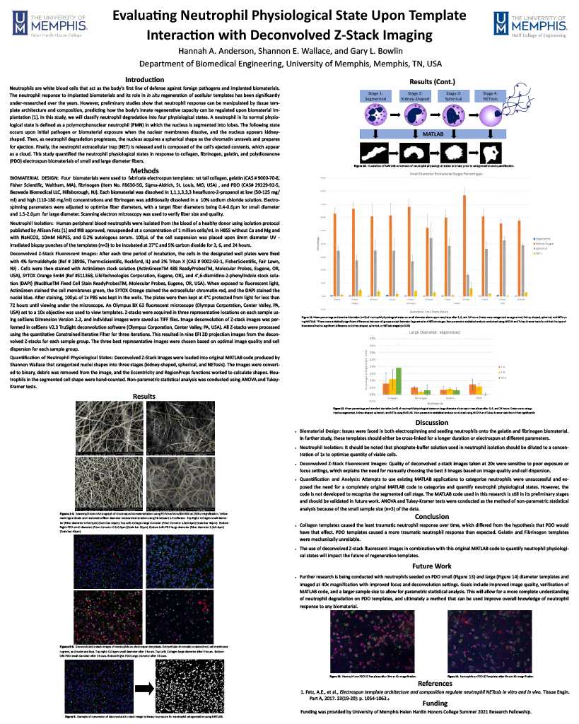
Evaluating Neutrophil Physiological State Upon Template Interaction with Deconvolved Z-Stack Imaging
Faculty Mentor: Gary Bowlin, Biomedical Engineering
Abstract: Our knowledge regarding neutrophil response to biomaterials is still limited. In this study, we classified neutrophil degradation into four physiological states, with the final stage being NETosis. Quantifications of these neutrophil physiological states were evaluated in response to collagen, fibrinogen, gelatin, and polydioxanone (PDO) electrospun templates with target diameters of 0.4-0.6 µm for small diameter and 1.5-2.0µm for large diameter. It was hypothesized that PDO would induce the least traumatic neutrophil response. The biomaterials were electrospun at fiber diameters 0.58 ± 0.2µm and 1.68 ± 0.2µm. Neutrophils’ in vitro responses were evaluated at 3, 6, and 24 hours. Deconvolved Z-stack fluorescent images were produced. Original MATLAB code was applied to images to quantify neutrophil states. Non-parametric statistical analysis was conducted. In this preliminary data (n=3), collagen electrospun biomaterial caused the least traumatic neutrophil response, while PDO templates caused a more traumatic neutrophil response than hypothesized. Gelatin and fibrinogen templates provided a mechanically unreliable response. This study was the first of its kind to combine this novel imaging technique in combination with original MATLAB code to quantify neutrophil states. In future research, larger sample sizes and parametric statistical analysis will produce meaningful data to impact the next generation of regeneration templates.
Sarah Coleman
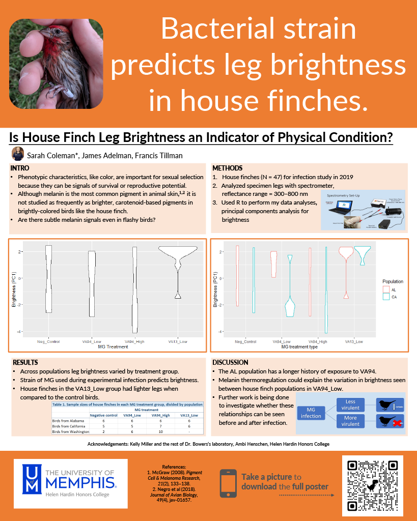
Is House Finch Leg Brightness an Indicator of Physical Condition?
Faculty Mentor: James S. Adelman, Biological Sciences
Abstract: Among various animals, mate choice is often associated with physical characteristics, such as coloration, which signal survival or reproductive potential. Although bright, flashy colors, like reds and yellows, are often studied, it remains unclear whether subtler colors, like browns and grays, also play a role. To determine if subtle coloration correlate with fitness in a species that also displays well-known flashy signals, I examined house finch (Haemorhous mexicanus) leg color after experimental infection with a bacterial pathogen, Mycoplasma gallisepticum, which causes seasonal epidemics in this species. I used spectrometry to determine whether leg brightness (visible and UV spectra) differed between finches inoculated with isolates of varying doses and virulence. Results indicated that birds infected with the more virulent isolate had lighter legs compared to the control birds. These results suggest that subtle grays and browns could indicate infection status, and therefore survival potential, in a species known to show signals using coloration. However, because these birds were tested after infection, further work is needed to investigate whether these same relationships signal fitness during infection, which better indicates survival probability. Nevertheless, my work suggests that subtle coloration could play role in signaling survival, with the potential to influence mate choice.

Juan Esparza
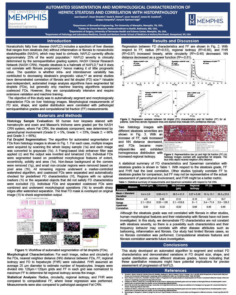
Automated Segmentation and Morphological Characterization of Hepatic Steatosis and Correlation with Histopathology
Faculty Mentor: Aaryani Tipirneni-Sajja, Biomedical Engineering
Abstract: Prevalence of non-alcoholic fatty liver disease (NAFLD) has increased to 25% of the world population, with one-third of NAFLD population progressed to irreversible non-alcoholic steatohepatitis (NASH). The current standard to assess NAFLD is liver biopsy, where pathologists use a semi-quantitative fat clinical research network (Fat CRN) visual grading system, which can have bias from inter-reader variability. To our knowledge, no study has described fat droplet (FD) characteristics and their relationship with pathologists’ grade, which would aid understand FDs’ changes and NASH progression to detect NAFLD at an early stage. In this study, a human cohort of NASH candidates were histopathologically graded by an experienced pathologist. FD automatic segmentation of FDs was performed and their morphology was described with radius, circularity, and distribution (nearest neighbor (NN) distance, regional anisotropy), and Fat CRN scores were predicted with FD to hepatocyte ratio (FHR, i.e., estimated affected hepatocytes) . Radius (R2=0.83,ρ=0.65), FHR (R2=0.87,ρ=0.85), and regional anisotropy (R2=0.66,ρ=0.69) had positive linear relationships and circularity (R2=0.48,ρ=-0.32) and NN distance (R2=0.81,ρ=-0.818) had negative relationships with FF. In conclusion, our study showed that size and distribution correlated well with pathologist’s grading, hence demonstrating that morphological characteristics might have potential implications for assessing disease progression.

Samantha Hall
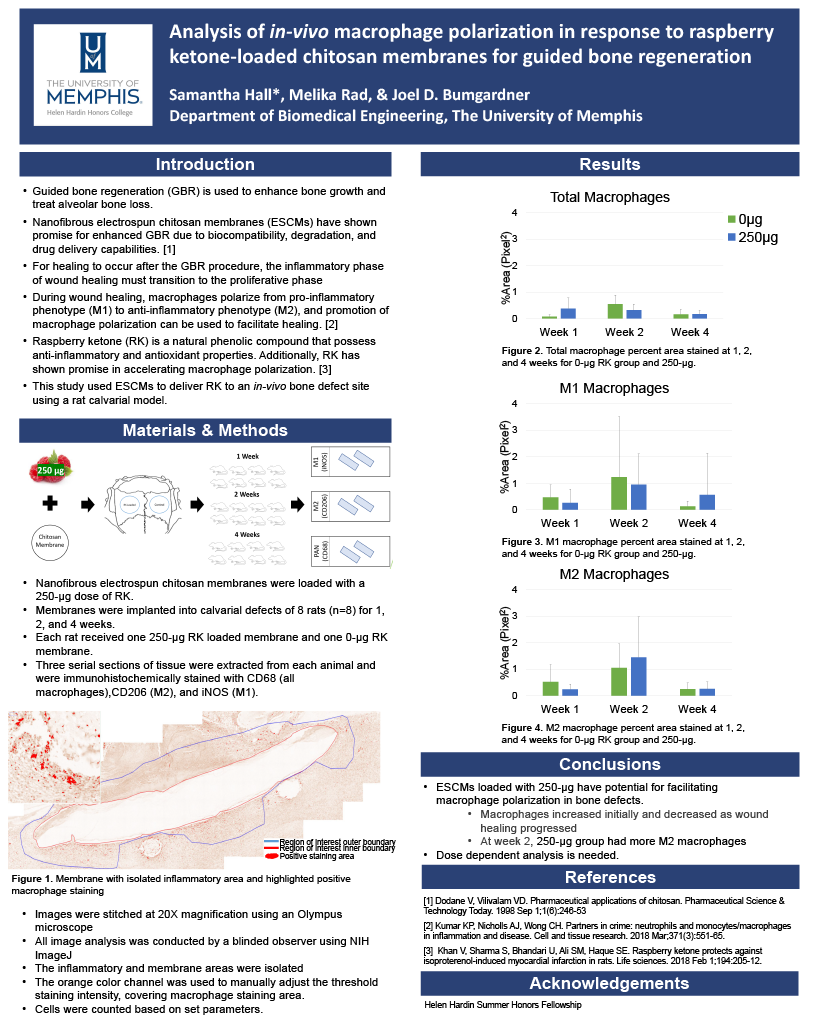
Analysis of in-vivo Macrophage Polarization in Response to Raspberry Ketone-loaded Chitosan Membranes for Guided Bone Regeneration
Faculty Mentor: Joel Bumgardner, Biomedical Engineering
Abstract: Guided bone regeneration (GBR) is used to enhance bone growth and treat alveolar bone loss. Nanofibrous electrospun membranes made from chitosan have shown promise for enhanced GBR. During wound healing, macrophages polarize along a spectrum from a pro-inflammatory phenotype (M1) to an anti-inflammatory phenotype (M2). A strategy that can be implemented to facilitate healing is the promotion of macrophage polarization. Raspberry ketone (RK) is a natural phenolic compound that possesses antioxidant and anti-inflammatory properties. In previous in-vitro research, RK has shown promise in accelerating macrophage polarization. In this study, electrospun chitosan membranes (ESCMs) were used to deliver RK to an in-vivo bone defect site using a rat calvarial model. ESCMs were loaded with 250 or 0 µg RK. Membranes from each treatment group were implanted into rat calvarial defects (n=8). Membranes and surrounding tissues were extracted in serial sections and immunohistochemically stained at 1, 2, and 4 weeks using individual markers for M1(iNOS), M2 (CD206), and total macrophages (CD68). Images of the stained tissues were obtained, and the percent-stained area was quantified using NIH ImageJ. Results indicated that ESCMs loaded with 250 µg RK have potential for facilitating macrophage polarization in bone defects. However, further dose dependent analysis is required.

Razan Sweileh
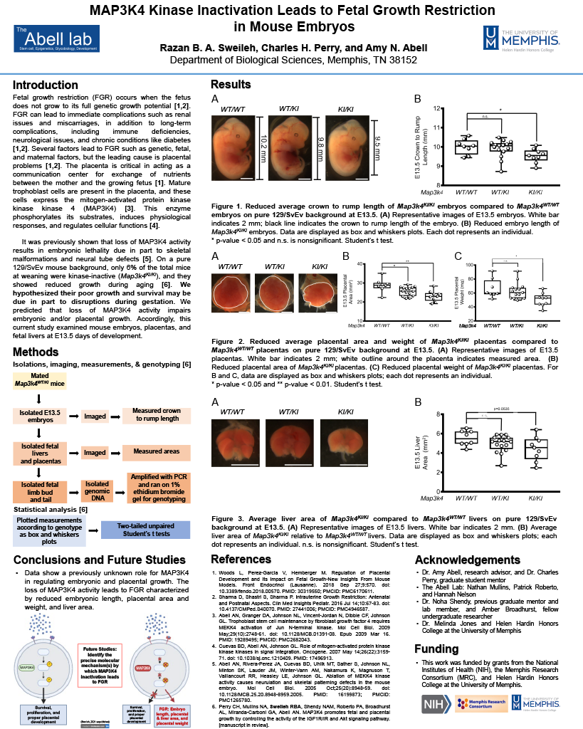
MAP3K4 Kinase Inactivation Leads to Fetal Growth Restriction in Mouse Embryos
Faculty Mentor: Amy N. Abell, Biological Sciences
Abstract: Fetal growth restriction (FGR) occurs when the fetus does not grow to its full, genetic potential. FGR causes life-long complications, including immune deficiencies and neurological issues. The leading cause of FGR is placental defects; the placenta is the communication channel between the mother and the growing fetus. The placenta is composed of mature trophoblast cells that highly express the mitogen-activated protein kinase kinase kinase 4 (MAP3K4). This enzyme phosphorylates its downstream substrates to regulate cellular functions and induce physiological responses for proper development. Inactivation of the Map3k4 gene results in embryonic lethality. Surviving kinase-inactive mice show reduced growth during aging. We hypothesized the complications in adults were due in part to disruptions during gestation. Subsequently, this current study examined embryonic length, liver area, and placental size in mouse embryos at 13.5 days of development. We found that inactive MAP3K4 embryos (Map3K4kIkI) had reduced embryonic length, liver area, and placental size when compared to wild-type Map3k4 embryos (Map3k4WT/WT). These findings indicate that MAP3K4 controls placental and embryonic growth, and loss of MAP3K4 activity leads to FGR. Future studies will focus on examining the placenta to understand the molecular mechanisms controlling the pathology of FGR.

Shannon Wallace
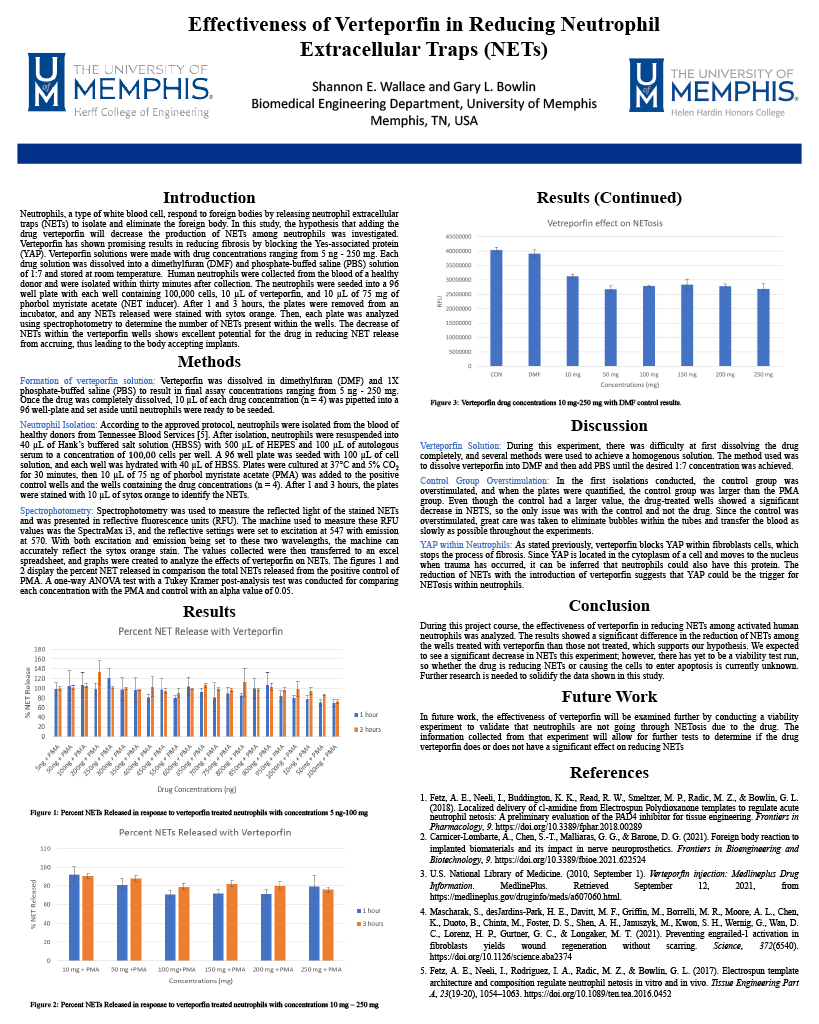
Effectiveness of Verteporfin in Reducing Neutrophil Extracellular Traps (NETs)
Faculty Mentor: Gary Bowlin, Biomedical Engineering
Abstract: Neutrophils, a type of white blood cell, respond to foreign bodies by releasing neutrophil extracellular traps (NETs) to isolate and eliminate the foreign body. In this study, the drug verteporfin was analyzed to determine its effects on NET release. Verteporfin has shown promising results in reducing fibrosis by blocking the YAP protein. Verteporfin solutions were made with drug concentrations ranging from 5 ng-200 mg. Each drug solution was dissolved into a dimethylfuran (DMF) and phosphate-buffed saline (PBS) solution of 1:7 and stored at room temperature. Human neutrophils were collected from the blood of a healthy donor and were isolated within thirty minutes after collection. The neutrophils were seeded into a 96 well plate with each well containing 100,00 cells, 10 µL of verteporfin, and 10 µL of 75 mg of phorbol myristate acetate (NET inducer). After 1 and 3 hours, the plates were removed from an incubator and any NETs released stained with sytox orange. Then, each plate was analyzed using spectrophotometry to determine the amount of NETs present within the wells. The decrease of NETs within the verteporfin wells shows great potential for the drug in reducing NET release from accruing, thus leading to the body accepting implants.